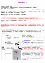
CASE환자의 MRI와 EEG 소견은
(1) precentral gyrus (2) middle frontal gyrus (3) cingulate gyrus 에 병변이 있는 것으로 판단되었다.
그리고 환자의 angiography 소견에서
(1) Anterior cerebral artery (2) Middle cerebral artery (3) Posterior communicating artery가 폐쇄되어 영상에서 보이지 않는 것으로 보였다. 이 혈관들이 분포하는 영역에
Development background
2. History of development
1960s
Discovery of colony-stimulating factors (CSF)
1980s
GM-CSF, M-CSF, G-CSF and multi-CSF
The human G-CSF was cloned
Usage for the treatment at first
1990s
first approval of Filgrastim by the US Food and Drug Administration
Launched Lenograstim
2000s
Launched PEG-filgrastim
1. Cerebrovascular Disease
(1) 정의
뇌혈관의 병적 변화에 의해서 나타나는 Brain의 이상상태를 의미하며 흔히 Stroke(뇌졸중)이라고도 한다. Cerebrovascular Accident(CVA)라고도 한다.
(2) 분류
Ischemic Stroke : 어떤 원인으로 Blood의 공급이 충분치 못할 때 발생 → Infarction
Hemorrhagic Stroke
⊙ Introduction
Postural tone는 중력에 대하여 일정한 높이를 지니고 있어야 한다. 정상인의 경우 움직임이 부드럽게 일어나려면 postural tone이 적절하게 조절 된다. 어떠한 방향으로 움직이든지 저항이 나타나지 아니하고 mobility를 유지하도록 한다. 그러나 중추신경계가 손상되었을 경우 이자세 tone에 이
1 개요
뇌는 신경계의 총수로 두개골 속에 있는 약 1500g 정도의 기관이다. 척수와 함께 중추신경계를 이루고 있다. 뇌는 척수처럼 반사의 중추의 역할을 하기도 하지만 부위에 따라 보다 고차원적인 기능의 여러 가지 활동을 한다. 중추신경계 중 척수가 가장 하위수준이며, 연수, 교, 중뇌, 시상하부,
Stem cell calssification
1. Based upon location
Embryonic stem cells
Somatic/ adult stem cells
2. Based upon function
Totipotent
Pluripotent
Multipotent
Unipotent
Reject Response
Traditional Immuno-repressents
Constant administration
Little engraftment after 3~4 weeks
Host immune system impaired
Vulnerable to Infection
↓Need brief immuno-re
Alzheimer’s Disease(AD)
퇴행성 뇌질환으로, 노화의 과정 속에서 뇌조직이 기능을 잃으면서 점차 정신 기능이 쇠퇴하는 병
노인에게 주로 나타나는 치매의 주요 원인 가운데 하나이며 병리조직학적으로는 뇌의 전반적인 위축, 뇌실의 확장, 신경섬유의 다발성 병변과 초로성 반점 등의 특징을 보임.
Alz
Mid-sagittal section
↓
Right hemisphere
↓
Anterior commissure & Posterior commissure 의 중간을 수직으로 section
↓
왼쪽사진-우반구의 뒷부분
오른쪽사진-우반구의 앞부분
Section 1
움푹패인 Lateral ventricle의 흔적
Corpus callosum
* 뇌의 관상단면 번호는 해당 순서의 단면이 아니라 일단 자른 14개의 단면 중에서
Thalamus의 lateral 경계를 이루는 것이 Stria Terminalis인데 여기를 따라 주행하는 vessel이 Thalamostriate Vein이다.
Interventricular foramen에서부터 opening of cerebral queduct까지 이어져서 third ventricle의 가쪽벽을 시상과 시상하부 구역으로 나눈다. (파란색)
Limbic lobe
Hippocampal formation
Amygdala
Septal Nuclei
Hypothalamus
Pa
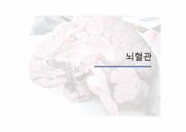
뇌의 혈액 공급 Vertebral a.와 internal carotid a.
Vertebral artery의 분포
: 뇌의 caudal 쪽 절반 마름뇌(Rhombencephalon)
중간뇌(Mesencephalon)
사이뇌(Diencephalon)의 꼬리쪽 부분
끝뇌(Telencephalon)의 occipital lobe과 temporal lobe의 basal portion
Internal carotid artery의 분포\
: 나머지 부분 Thalamus의 앞쪽 절반
줄무늬체(corpus striatum)